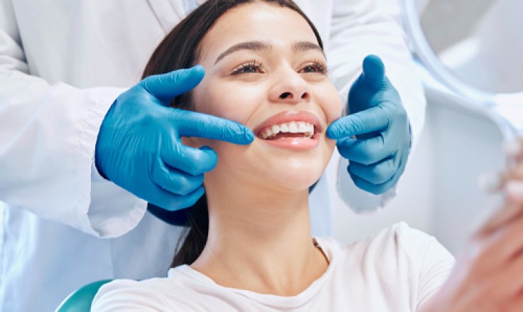
Introduction
3D imaging in dental treatments has transformed the way dentists diagnose, plan and perform procedures. The technology provides detailed three-dimensional views of teeth, bone and surrounding structures. Traditional two-dimensional X-rays offer limited perspective. 3D imaging in dental treatments reveals far more anatomical detail. Many modern dental clinics now use this technology as standard practice. Patients benefit from more accurate diagnosis and more predictable treatment outcomes. Understanding the role of this technique in dental treatments helps patients appreciate the advantages of clinics that invest in this advanced technology. This article explains how the technology works, its key applications and the benefits it delivers for patients.
What Is 3D Imaging in Dental Treatments?
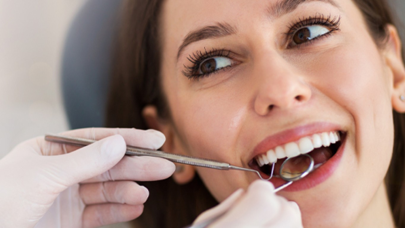
3D imaging in dental treatments refers to advanced scanning technology that creates three-dimensional representations of oral and facial structures. The most common technology is cone beam computed tomography, known as CBCT. The scanner captures hundreds of images from different angles as it rotates around the patient’s head. Specialised software reconstructs these images into a detailed three-dimensional model. The resulting scan shows teeth, bone, nerves, sinuses and soft tissues in three dimensions. The technology provides significantly more information than traditional dental X-rays. 3D imaging in dental treatments allows dentists to view structures from any angle. This comprehensive visualisation supports more accurate diagnosis and more precise treatment planning.
How 3D Imaging in Dental Treatments Works
Scanning process for 3D imaging in dental treatments is quick and comfortable. Patient stands or sits in the scanner. The machine rotates around the head over ten to forty seconds. The scan involves much lower radiation than a medical CT scan. Specialised software processes the captured data immediately. The dentist can view the three-dimensional model within minutes. The model can be rotated, sectioned and measured using digital tools. Cross-sectional views reveal internal structures. Virtual models show bone density and volume accurately. The technology allows precise measurements for implant placement. Multiple views can be generated from a single scan, eliminating the need for repeated imaging.
3D Imaging in Dental Treatments for Implant Planning
Implant planning represents one of the most valuable applications of 3D imaging in dental treatments. The technology reveals exact bone dimensions where implants will be placed. Height, width and density of available bone are measured precisely. The position of nearby nerves and sinuses is identified accurately. This information allows the dentist to plan implant placement with millimetre precision. Guided surgery using 3D printed drill guides can be created from the imaging data. These guides ensure implants are placed exactly where planned. The technology reduces surgical risks and improves outcomes for dental implant patients. Complex cases including full arch restorations benefit particularly from the detailed planning that this particular technique provides.
3D Imaging for Orthodontic Treatment Planning
Orthodontic treatment benefits significantly from 3D imaging in dental treatments. The technology reveals the relationship between teeth, roots and surrounding bone in three dimensions. Hidden teeth, unusual root positions and bone anomalies are identified clearly. Treatment planning for clear aligners uses digital models created from 3D scans. The technology allows virtual simulation of tooth movements before treatment begins. Patients can preview their expected results digitally. Complex orthodontic cases requiring precise movement sequences benefit from detailed imaging. The combination of surface scanning and CBCT creates comprehensive digital models. These models support the most accurate orthodontic treatment planning available in modern dentistry.
3D Imaging in Dental Treatments for Endodontics
Root canal treatment is another area enhanced by 3D imaging in dental treatments. The technology reveals root canal anatomy in detail. Additional or curved canals that might be missed on traditional X-rays become visible. The extent of infection around the root tip can be assessed accurately. This information helps endodontists plan more effective treatment. Failed previous root canal treatments can be evaluated in three dimensions. Cracks, fractures and resorption are identified more reliably. The technology improves diagnosis of complex cases. Treatment outcomes improve when anatomy is fully understood before the procedure begins. The technique has become particularly valuable for challenging endodontic cases.
Smile Design and 3D Imaging
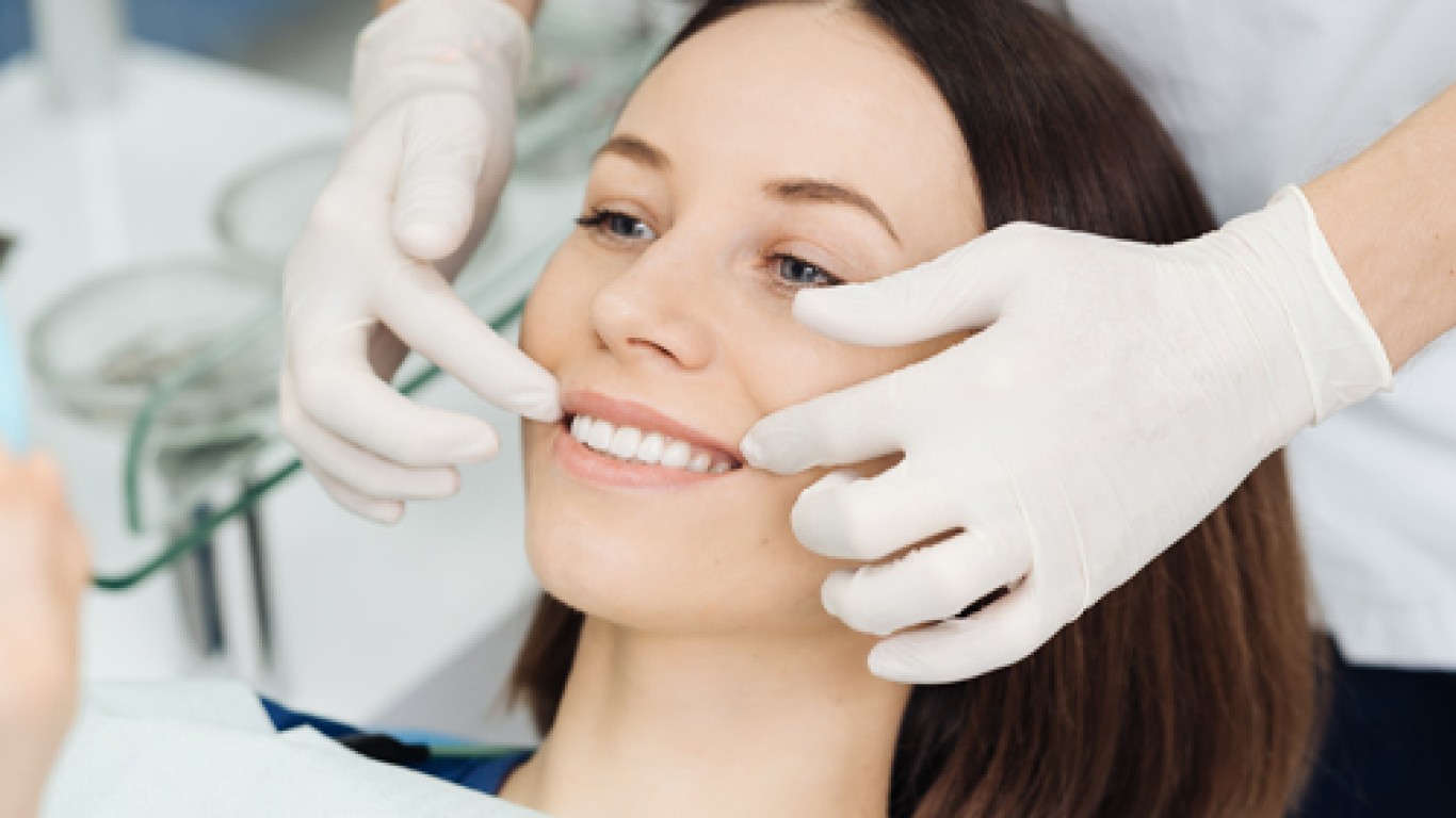
Cosmetic dentistry benefits from 3D imaging in dental treatments through digital smile design capabilities. Surface scanners create detailed three-dimensional models of existing teeth. The digital model allows the dentist to design restorations precisely. Veneers, crowns and bridges are designed digitally before fabrication. Patients can preview their new smile on screen. Adjustments are made digitally until the desired appearance is achieved. CAD/CAM technology uses the digital design to manufacture restorations accurately. The fit is typically superior to traditional impression-based methods. 3D imaging in dental treatments streamlines the process and improves outcomes for cosmetic dental patients. The precision of digital planning produces more predictable and satisfying aesthetic results.
Benefits of 3D Imaging for Patients
3D imaging in dental treatments offers multiple benefits for patients. Diagnosis is more accurate because the technology reveals structures invisible on traditional X-rays. Treatment planning is more precise because exact measurements guide clinical decisions. Surgical procedures are safer because critical anatomical structures are identified in advance. Treatment outcomes are more predictable because planning accounts for individual anatomy in detail. The scanning process is quick, comfortable and non-invasive. Digital records can be stored and compared over time. Communication between dental specialists improves when all providers can view the same detailed imaging. Patients benefit from understanding their condition better through visual three-dimensional models.
3D Imaging in Dental Treatments vs Traditional X-Rays
3D imaging in dental treatments differs from traditional X-rays in several important ways. Traditional dental X-rays produce two-dimensional images. Structures overlap, making interpretation challenging. Small details can be missed. The technique creates a complete three-dimensional model. Each structure is visible separately from every angle. Bone volume and density are measured accurately. Nerve pathways are traced precisely. The technology provides significantly more diagnostic information. Traditional X-rays remain useful for routine screening and simple assessments. 3D imaging is indicated for complex treatment planning, implant placement, orthodontic assessment and evaluation of difficult cases. The two technologies complement each other in comprehensive dental care.
Conclusion
3D imaging in dental treatments has transformed modern dentistry through more accurate diagnosis and precise treatment planning. The technology benefits implant placement, orthodontics, endodontics and cosmetic dental procedures. Patients enjoy safer procedures, more predictable outcomes and the ability to preview results before treatment begins. The scanning process is quick, comfortable and involves minimal radiation. Choosing a clinic that utilises this technique in dental treatments supports the best possible clinical outcomes. The technology represents a significant advancement over traditional two-dimensional X-rays. Professional consultation at a clinic equipped with this technology ensures each patient receives the most thorough assessment and personalised treatment plan available. Continued advancements in imaging technology are expected to further enhance diagnostic accuracy and patient experience.
To understand more about the technology and treatment planning process visit the ACIBADEM Beauty Center Dental Treatments page.
Frequently Asked Questions
Advanced scanning that creates three-dimensional models of teeth, bone and oral structures.
Yes, the radiation exposure is comparable to a few traditional dental X-rays.
Not always; it is most valuable for implants, orthodontics and complex cases.
The scan typically takes ten to forty seconds.
No, the scan is completely comfortable and non-invasive.