Introduction
The position of the upper eyelid plays an important role in both vision and overall facial balance. Even a small change in eyelid height can influence how alert or fatigued someone appears. When the upper lid droops lower than normal, it is known as ptosis of the eyelid. This condition may affect one or both eyes and can vary in severity. In mild cases, it causes subtle asymmetry that may be mostly aesthetic. In more pronounced cases, it can interfere with vision and daily activities. Understanding the causes and treatment options helps patients seek appropriate care confidently and at the right time.
What Is Ptosis of the Eyelid?
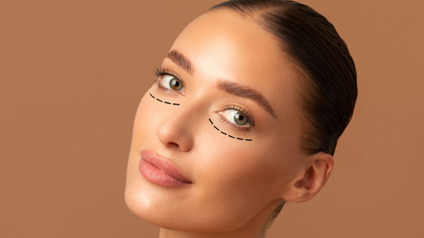
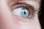
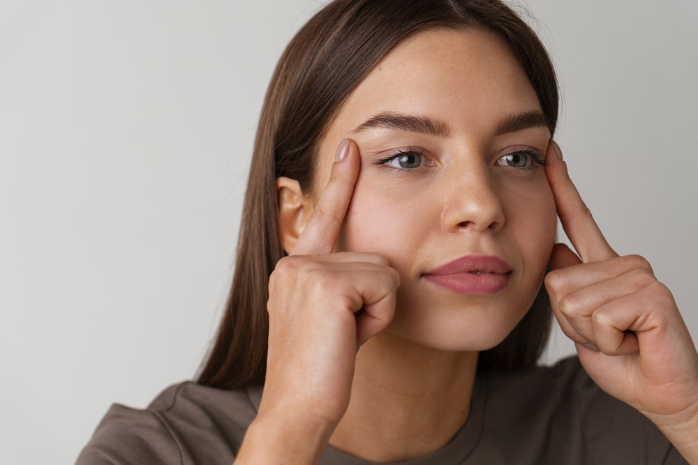
Ptosis of the eyelid refers to a lowering of the upper eyelid margin beyond its normal anatomical position. The eyelid may partially cover the pupil, depending on severity and muscle strength. This occurs when the muscle responsible for lifting the lid weakens, stretches or detaches slightly. The levator muscle plays a central role in eyelid elevation and blink coordination. When its function declines, the lid position changes and symmetry may be lost. Diagnosis requires careful examination by a specialist to determine the degree of drooping and its functional impact.
Ptosis of the Eyelid: Congenital Ptosis
Some individuals are born with ptosis of the eyelid, a condition known as congenital ptosis. It usually results from underdevelopment or poor function of the levator muscle. The condition may affect one eye more than the other, creating visible imbalance. In children, significant drooping can interfere with visual development if the pupil is obstructed. Early assessment is important to prevent long term visual complications. Treatment timing depends on severity, functional limitation and the risk of developmental issues.
Ptosis of the Eyelid: Acquired Ptosis
Acquired ptosis of the eyelid develops later in life and is more common in adults. Age-related muscle stretching is the most frequent cause. Over time, the levator muscle may weaken gradually due to natural tissue changes. Trauma, previous eye surgery or long term contact lens use may also contribute. Certain neurological or muscular conditions can affect nerve or muscle control. Identifying the underlying cause helps guide treatment decisions and ensures appropriate correction.
Symptoms Beyond Drooping
The most obvious sign of ptosis of the eyelid is visible drooping of the upper lid. However, other symptoms may also appear and affect comfort. Some individuals raise their eyebrows frequently to compensate for the lowered lid position. This repeated action can cause forehead tension and fatigue over time. Head tilting backward may occur to improve visual access. In more severe cases, upper field vision becomes limited and daily activities may feel strained.
Differences Between Mild, Moderate and Severe Cases
Ptosis of the eyelid can vary significantly in severity.
- In mild cases, the droop may be barely noticeable and primarily aesthetic.
- Moderate cases may partially cover the pupil and create visible asymmetry.
- Severe ptosis can obstruct a significant portion of vision.
- The degree of drooping influences both treatment timing and technique.
Careful classification helps guide whether observation or surgical correction is appropriate. Accurate grading ensures realistic expectations and proportionate intervention.
Impact of Ptosis of the Eyelid on Children and Visual Development
When ptosis of the eyelid occurs in children, early assessment becomes particularly important. If the eyelid covers the pupil consistently, visual development may be affected. The brain may favour the unobstructed eye, potentially leading to imbalance. Prompt evaluation helps determine whether early surgical correction is necessary. In mild congenital cases, monitoring may be appropriate. Protecting long term visual clarity is a central priority in paediatric assessment.
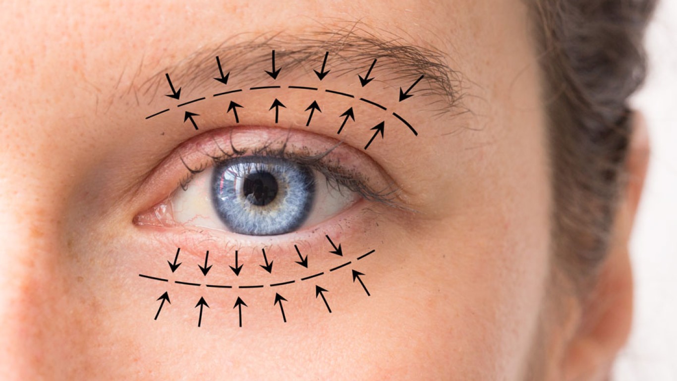
How Ptosis of the Eyelid Is Diagnosed
Diagnosis begins with a comprehensive eye and eyelid examination.
- Eyelid height, margin position and symmetry are measured carefully.
- Muscle strength testing evaluates levator function through guided eye movements.
- Visual field testing may assess functional impairment caused by drooping.
- Brow position is also evaluated, as it influences perceived eyelid height.
A clear diagnosis determines whether ptosis of the eyelid is mild, moderate or advanced. Accurate assessment guides personalised treatment planning.
The Role of Brow Compensation
Many individuals with ptosis unconsciously lift their eyebrows to improve vision. This compensation can temporarily raise the eyelid position. However, constant brow elevation may cause forehead fatigue and tension. Over time, horizontal forehead lines may deepen due to repetitive lifting. Treating ptosis of the eyelid reduces the need for this muscular compensation. Restoring proper lid height allows the forehead to relax naturally.
Distinguishing Ptosis of the Eyelid From Dermatochalasis
Ptosis of the eyelid is sometimes confused with dermatochalasis, which refers to excess upper eyelid skin. While both conditions create heaviness, their causes differ. Dermatochalasis involves loose surface skin, whereas ptosis relates to muscle weakness. In some cases, both conditions occur together. Accurate diagnosis determines whether muscle tightening, skin removal or combined treatment is required. Proper differentiation ensures the correct procedure is selected.
Non Surgical Treatment Options
In mild cases, non surgical approaches may be discussed as temporary measures. Specialised glasses with eyelid crutches can support lid elevation mechanically. These are typically short term solutions rather than permanent correction, and don’t address the underlying muscle weakness. Observation may be appropriate when functional impact is minimal. However, long-term correction for ptosis of the eyelid often requires surgical adjustment for reliable results.
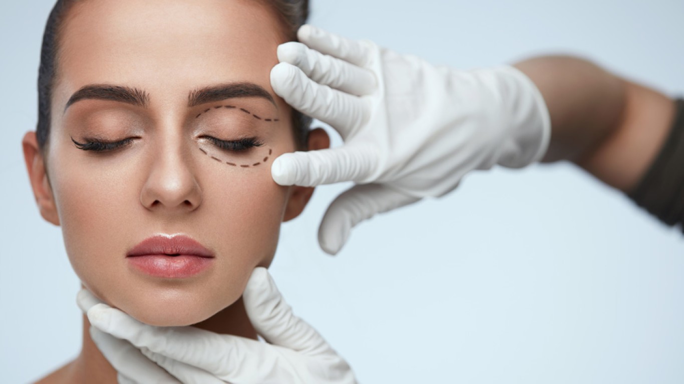
Surgical Treatment for Ptosis of the Eyelid
Surgery remains the most effective treatment for ptosis of the eyelid when functional or aesthetic concerns are significant. The procedure involves tightening, shortening or repositioning the levator muscle to restore lift. A small incision is usually placed within the natural eyelid crease to allow discreet healing. In some cases, alternative muscle support techniques are used when levator strength is limited. The goal is to restore natural eyelid height, symmetry and smooth movement.
Internal Versus External Surgical Techniques
There are different surgical approaches to correcting ptosis of the eyelid. External approaches involve a small incision in the natural crease. Internal techniques may be performed from the underside of the eyelid in selected cases. The choice depends on muscle strength and eyelid anatomy. Each method aims to restore lift while preserving natural contour, and technique selection is tailored carefully to individual needs.
Conclusion
Ptosis of the eyelid is a condition that affects both appearance and function. Identifying the underlying cause is essential before selecting treatment. While mild cases may be monitored, surgery often provides lasting and meaningful correction. With accurate diagnosis and careful technique, eyelid position can be restored effectively and proportionately.
If you’re interested in finding out more about eyelid surgery, visit the ACIBADEM Beauty Center blepharoplasty page.
Frequently Asked Questions
It is usually caused by muscle weakness or stretching.
No, it can develop later in life.
Yes, severe cases may limit upper field vision.
Surgery is the most effective long term option.
Most swelling improves within two weeks.